

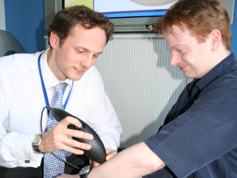
Astron Clinica SIAscope V skin imaging camera
SIAscopy skin imaging
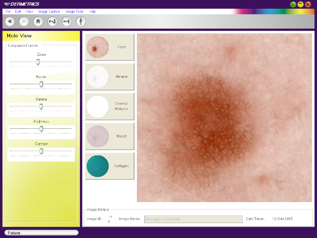
Skin cancer kills 60,000 every year, but is very treatable when detected at an early stage. SIAscopy—Spectrophotometric Intracutaneous Analysis, a technology developed by Symon Cotton on the basis of his PhD-thesis—has been shown to facilitate diagnosis. SIAscopy is based on multispectral imaging, allowing visualisations of the distribution of melanin (pigment), blood and collagen in the skin.
SIAscans are made with a dedicated camera and image processing software. Astron Clinica had been selling previous model SIAscopes in limited numbers for a few years, but were looking at drastically reducing the hardware cost, weight and size while increasing image quality and improving ease of use. The hardware cost reduction would make it possible for them to move to a subscription business model.
“The fact that we could take an early model to take skin images during a meeting with our investors convinced them that our development plans and cost targets were realistic, and that the new SIAscope V would be ready in time”
Concept design
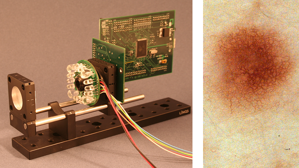
We came up with a number of possible camera concepts all based around the use of a 3Mpixel consumer-grade imager chip which would reduce the hardware cost by over 90%, allowing Astron Clinica to switch their business model from hardware sales to software subscriptions. Adding high-CRI white LEDs to the device also improved the dermatoscopic images.
The SIAscope V uses a USB 2.0 connection to a host PC to send images and draw power. A capacitor buffers power in the handset as a 1-second SIAscopy image sequence (2 images each flashing white, red, green, blue and infrared high-power LEDs) sinks over 500 mA. The white LED is continously lit at lower current to produce a “viewfinder” image stream.
Engineering
We specified, designed and prototyped all electronic circuits, optical components and mechanical parts from the ground up, taking into account the intended production volumes of upper hundreds to lower thousands per year. Prototypes were tested and refined in trials with nurses and dermatologists, which led to a decision to upgrade the initial lens specification—the increased value of better image quality more than offset the additional unit manufacturing cost.
Manufacture and sales
We assisted Astron Clinica to transfer our design to a local manufacturing partner. The SIAscope V was introduced in March 2006 in conjunction with the DERMETRICS imaging suite for clinical screening of skin lesions. The SIAscopy IP was later acquired by MedX Health.
The same device also found application in the cosmetics sector under the Cosmetrics as well as Olay’s BeauVisage brands.